Spinal Neurology and Manual Therapy
(& not only...)

  Вертеброневрология и мануальная терапия
(& не только...)


menu

Коричневый цвет - русская версия
Green Color - English version


Клиника восстановления здоровья
Body Rehabilitation Clinic
 

Виды лечения - Treatment Modalities

****** 

Testimonials

****** 


Body Rehabilitation Clinic
(AMTE Site)


Treatment Types

****** 


Американский Фонд изучения позвоночной неврологии
Orthopedic Neurology Research Fund
 

Цель Фонда - Fund's Goal

****** 

From Board Directory

****** 

Методические пособия

****** 

Учебные пособия

****** 

Об авторе

****** 

About the Author

****** 

Proceedings. Publications

****** 

Message to Colleagues

****** 


Невропатология и лечение межпозвонкового остеохондроза
Intervertebral Osteochondrosis' Neuropathology and Treatment
 

Как расставаться с хронической болью? (Беседа о восстановлении здоровья)

****** 

Повсеместные боли

****** 

Малоизвестная в США ортопедическая неврология

****** 

Less-known Spinal Neurology

****** 

Развитие отечественной вертеброневрологии

****** 

Нейрохирургия остеохондроза

****** 

Проблема века или вечная проблема?!

****** 

Сомнительные подходы в вертеброневрологии и мануальной терапии

****** 

Отечественные черты мануальной терапии

****** 

Вазодистонии и ишемии

****** 

Радикуло-миелоишемия

****** 

Межпозвонковый остеохондроз в Евразии и Америке

****** 

Полвека остеохондроза

****** 

Бенефиты манипуломании

****** 

Линия отчуждения

****** 

Медицинские сюрпризы

****** 

"Клиническая пропедевтика мануальной терапии" Монография

****** 

Отзыв на монографию

****** 

Своеобразие текущего момента

****** 

В защиту суверенитета вертеброневрологии

****** 

О создателе клинической дисциплины
About the Founder
 

Ближайший взгляд

****** 

От последователей

****** 

Патриарх неврологии

****** 

Феномен Якова Попелянского

****** 

Об Отце и его Деле

****** 

Казань - Сиэтл

****** 

Он опережал время

****** 

Памяти Я. Ю. Попелянского

****** 

In memory of Professor Yakov Popelyanskiy

****** 

Основополагающие первоисточники - Spinal Neurology Textbooks

****** 

Руководство и монографии по неврологии

****** 

Some Articles

****** 

Другие книги профессора Я. Попелянского

****** 

Opinions of World prominent Specialists

****** 

Статья из Неврологического Журнала

****** 

Учителю

****** 

Идеи Проф. Я. Ю. Попелянского в Америке

****** 

Гений и злодейство

****** 

Последнее интервью

****** 

Я люблю Вас живого

****** 

Научная биография

****** 

"Запоздалое открытие"

****** 

Я. Ю. и Политбюро

****** 

Компетенция и некомпетенция

****** 

О Солженицыне

****** 

Антисемитизм глазами невропатолога

****** 

Медицина в США глазами иммигранта-врача

****** 

Я. Ю. и поэзия

****** 

Неопубликованное

****** 

Обнаруженное

****** 


Хобби и отдых
Hobby and Entertainment
 

Обращение

****** 

Дисбаланс

****** 

Бальзам прошлого

****** 

Вне расписания

****** 

Уроки балкарского...

****** 

Брызги шампанского...

****** 

Выплывшее

****** 

Непредвиденное

****** 

О названии

****** 

Нескромная прелесть провинции

****** 

Вокруг "Возвращения"

****** 

Навеянное

****** 

Откровение

****** 

Наблюдаемое

****** 

Лицемерие и ханжество

****** 

Вокруг да около

****** 

Выборы

****** 

Конец света

****** 

Трагическое разочарование

****** 

Обновленное прояснение

****** 

Холодная гражданская война

****** 

Фашизм и прогресс

****** 


Vertebroneurology, orthopedic neurology, osteochondrosis, column, vertebral disc, syndromes, backache, neckpain.


Full text of the Manual "Vertebrogenic Diseases of the Nervous System "  - waiting for translation.


List of Publications - waiting for translation.


Dear colleagues, neurologists, neurosurgeons, orthopedists, radiologists, rheumatologists, physiotherapists, rehabilitologists and specialists of manual medicine, all those of you who became specialists in their respective fields after learning the basics of vertebroneurology!

I hope that this manual and, specifically, its selected chapters relevant to this discipline and presented below, will be interesting to otiatrists and especially to otoneurologists and internists.

Russian readers are familiar with this problem through my manuals as well as through monographies of my collaborators.

Right now, I would ask my foreign colleagues who are about to start reading this material about only one essential issue:  

Do not let yourself be guided by preconception!


The author of this site, Yakov Yu. Popelyanskiy was chair of the departments of neurology in several Russian universities for 40 years.

He was a professor, an acclaimed Russian scientist, a honorary academician of Euro-Asian Academy of Medical Sciences.

Prior to December 2000, he was the head of the All-Russian Center of Vertebroneurology in Kazan, Russia.


Yakov Yu. Popelyanskiy


(1918 - 2003)

LESS-KNOWN VERTEBRONEUROLOGY

The Russian neurologists demonstrated that at "backache", "neckpains" etc. are absoleted not only the statements of "Radiculitis", "Scialgia", but and the statement about a compressio of roots as ostensibly the main neurologic display of a discs hernia.

They described a great number of another syndromes - reflexive and myo-adaptive, which make the contents of a new medical discipline - vertebroneurology or orthopedic neurology .


Due to well-known difficulties of Russian medicine during the last 10 years of isolation, since the 60s, it has fallen behind the world medicine in the case of vertebroneurology too, and especially if the problem of discopathy is concerned. Such a gap was filled by the effort of Novokuznetsk and Kazan neurologists, neurosurgeons and orthopedists of a number of cities of Russia. That happened owing to the fact that there existed auspicious ideological prerequisites in the sphere of so-called neurism (Sechenov, Pavlov, Anochin, Speransky, Bernstein).

Therefore being behind their foreign colleagues in surgical maintenance, being devoid of necessary professional contacts, necessary equipment and financial maintenance, Russian scientists concentrated their efforts on clinical-neurophisiological and neuromorphological aspects of neurovertebrology.

With the purpose of the development of neurological aspect of the problem there have been obtained modern data unknown to not Russian-language readers.

The scientists have proved that the mechanical factor of radicle compression by disc hernia is only the simplest and partial (somehow primitive) part of the problem.

The All-Russian center of this medical discipline (vertebroneurology) at the Ministry of Health of Russia has been headed since 1978 just by the author of this report.

It turned out that radicle compression of the spinal cord or vessels occurs more rarely than among one third of patients with vertebrogenic diseases of the nervous system.

The majority of these patients suffer not from the radicle compression but from reflexive processes and reflexive syndromes. In response to lig. longitudinale posterior irritation by disc hernia or by other vertebral structures, pathological impulses are transferred through the intact segmental nervous apparatus and cause a reflexive spasm of cross-striatus muscles (paravertebral, membrum superior et inferior and cranial). They are muscle-tonic reflexive syndromes ( Fig. 1 ).

Fig. 1

The same are responses of smooth muscles of the support-motoric apparatus and other organs. They are reflexive neuro-vessel syndromes. The same are reflexive syndromes being realized not only in micro- but in macro- intervals of time as well; according to Speransky they are painful neuro-dystrophic syndromes.

They are the third variant of reflexive neuro-dystrophic syndromes.

They are all lesions of somatic structures, first of all of support-motoric apparatus.

Muscle contractions and dystrophy affect the deformation not only of the vertebral column, but extremities as well. Consequently secondary nidi of irritation appear, which cause new reflexive disturbances of the process of adaptation. They are myo-adaptive syndromes.

Therefore, our classification, beginning from 1962-1973 can be presented as follows (with some variations) it is considered to be an official one in Russia.

Without dwelling on the relatively simple question of compression syndromes (as they are presented quite well in the literature of West Europe and the USA) reflective syndromes of osteochondrosis will be briefly stated.

In Russia there is used the term "osteochondrosis" suggested by Hildebrandt in 1933.

It reflects the state of initiating dystrophic nidus - pulpose complex (nucleus) - chondros and osteon - of adjacent vertebrae. It is a serious disease. Another term, but disorienting one - spondylosis - is used in Great Britain. The later is not an essential pathology, not a lesion, but a change. It is an age, almost physiological (and more often age) bone adaptation to adjacent discs. That is a simplified observation of terminological point.

Some syndromes can be presented schematically and step-by-step as following:

Syndrome of a. vertebralis.

It can be caused by a mechanical influence of bone accretion (more often by proc. uncovertebralis) on the sympathetic plexus of vertebral artery.

Mechanism of reflexive variant: irritatio of receptors of n. vertebralis -> efferente fibers of n. vertebralis -> gl. stellatum -> efferente fibers of n. vertebralis -> gl. gl. spinales -> segmentale apparatus -> r. r. communicantes albi -> gl. gl. sympathicus cervicale -> sympaticale efferents of vessels of cranii (including a. auditiva interna) and cerebri.

Clinical pictures (they are quite well presented in the above-mentioned literature): cranialgia, cochle-vestibulare disturbances, tenderness of point of a. vertebralis.

We distinguish between functional and organic forms of the syndrome.

Cranialgia vertebrogenica sclerotomica and cranialgia vertebrogenica of strain.

Pathologically deformed cervical vertebral structures (including cranio-vertebral) are sources of irritation.

Painful irritation of the first variant according to sclerotomic, usually asymmetrical and little-depending on weather and emotion factors.

It is caused by jerky movements of the head and other mechanical influences. Strain-variant - according to all rules of the given type of cranialgia + dependence on the movements of the head.

Syndrome of m. obliaus inferior.

This muscle is an intervertebral analogue of rotatory muscles on the level of C I-II .

The muscle which is quite essential for the cranio-cervical movements increases many times more its volume in ontogenesis than other muscles.

During the reflexive contraction it sets its rotatory subluxatio C I-II .

When being congential or acquired defectiveness of this area the muscle easily reacts by reflexive contraction.

There appear local and sclerotomic pains - tenderness, torticollis (with the following feedback; output, response, into the occiput).

As over it n. occipitalis is bent, there can be signs of abaissement from the sight of this nerve trunk.

Cervicalgia (acuta, subacuta, chronica).

As well as dorso- and lumbalgia cervicalgia refers to the vertebral syndromes (in contradistinction to cervico- and pelviomembral syndromes).

It appears as a sclerotomic pain due to the irritation of receptors of n.synuvertebralis in lig. longitudinale  of column. It is in the whole a discogene component of the syndrome.

An important component of the syndrome is myotonic, that is a reflexive spasm of paravertebral muscles of the neck.

While the irritation is asymmetrical there appears a reflexive torticollis with a corresponding limit of movements of the head, tenderness of muscles and interosseus chords.

If there prevail dystrophic lesions of joint capsule (cervico-spondyloperiarthrosis) there capsules are painful.

Scalenus-anterior syndrome.

This well-known syndrome most often displays itself as a reflexive feed-back (output, response) of the indicated muscle: spasm, defance, contracture to pathology of neck discs.

Therefore reflexive displays appear, such as a contraction and tenderness of the muscle when movements in the neck section and when irritation of the source of the reflex take place.

Secondary compression displays are stimulated for compression of the lower trunk of plexus brachialis - a picture of irritatio et abaissment in region of intervation of the n. ulnaris.

Scalenus-media syndrome.

There is an analogous picture of reflexive displays + secondary compressive lesions. The muscle provides a pathological influence on a. transversa colli.

As its descending branch supplies the muscles of the scapula, suprashoulder,  a corresponding compressive and reflexive ischemia displays itself by painful ("neuralgic") stage of the disease. We consider this pain in the scapula section to be an analogue of the pain in myocardium.

The muscle influences n. thoracalis longus as well - and consequently - myotrophy of the muscle of the scapula (especially m. serratus anterior). From this the amyotrophic stage follows.

That is, in our view, the mechanism of neuralgic amyotrophy of Personage - Turner.

Naturally, the source of irritation can be not only the vertebral column.

Periarthrosis humero-scapularis.

This well known for orthopedists picture very often occurs "cervico-genic" reflexive.

Reflexive shortening of adductors of shoulder (m.m. pectoraiis and teres mayor) limits the movement of the shoulder aside, later reflexive neuro-dystrophic disturbances and in connective tissues of the elements of periarticulare tissues ( Fig.2 ):


Fig. 2

The patient P. Volume of possible active locomotions by a dextral arm in a frontal plane:
a - up to a novocainisation of a cervical intervertebral disc;
b
- straight away after a novocainisation of a disc.

Naturally, the source of irritation can be not only tissues of the vertebral column.

The prevalence of reflexive muscle-tonic and neuro-dystrophic disturbances in the greater pectoralis muscle can be often a cause of pseudo-cardiac syndrome of the front side of the thorax; the prevalence of muscle-tonic reaction in the minor side of the thorax - is a cause of the compression of the brachial plexus and axial artery, i.e. so-called "Saturday" paralysis or "police" paralysis of the muscles of the hand.

Dorsalgia.

Dorsalgia as well as pectalgia very often occurs as a consequence of dystrophic changes in the thorax part of the vertebral column.

However, due to the fixed state of this part, usually not osteochondrosis and joints have a clinical meaning, but heads and prominences of the rib, that is their arthroso- periarthrosis.

Palpation always allows to define lesioned level. The secondary neuropathy of the intercostal nerve - that is a rare find.

Lumbalgia.

Lumbalgia - acuta (lumbago), subacuta, chronica. The mechanism of the origin is considered above in the case of "cervicalgia".

Since kyphosation provides decompressive increase of sagittal diameter of the vertebral canal, during lumbalgia such a pose is formed. Not rarely there appears scoliosis.

It is formed by local muscles of vertebral segment - m. m. intertrasversarii, rotatores.

The tension of paravertebral muccles, especially m. m. multifidus is clearly defined by sight and by palpation.

When standing they are relaxed (the lardotic pose is achieved by the gravitation and not by the activity of the muscles).

When bending forward a little they are tensed. During the lumbar osteochondrosis these muscles are already tensed in the state of relaxation and do not relax when beading forward for a long time or when standing on one leg ("sign of ipsilateral strain") or other movement of the leg.

If reflexive miofixation of lesioned vertebral segment is sufficient a movement in the hip joint (sign of Lassegue) a disc is not traumatized and pain in the lumbar does not appear.

As it was mentioned above, all vertebrogene syndromes are divided according to the topical principle schematically into vertebral, ventrales (cervico-, dorso-, and himbalgia; here is possible to classify and coccygodynia) and  extravertebrales.

Thus, for example, extravertebral syndromes m. scalenes are not only extravertebrales, but vertebrates as well, since these muscles are joined by one of the tags to the vertebral columns.

That can be applied to the syndrome of the pear-shaped muscle as well.

Among pelviomembrale syndromes there are the following most often met syndromes:

Syndromes of pelviale bottom.

It is constituted of muscles (levator ani, coccygeus, gemelli, piriformis) and chords (first of all, sacrotuberale, sacrospinale).

Reflexive strain of these muscles and reflexive dystrophic disturbances of connective tissue structures of chords and muscles - that is a source of pain and deformations in this field.

In its turn, the shortening of m. piriformis and dystrophy of bottom chords causes compression neuropathy of n. pudendus.

So called, coccygodynia is more often caused not by a trauma of the osteale, but by muscular-tonic and neurodystrophic disturbances.

Summation of impulses from pathologic organs exhibited by pains and tenderness. Spina ischiadica is painful in 100%.

There are marked neuravasculare disturbances in the region of perineum and by signs of abaissement from the part of n. pudendus.

Piriformis syndrome.

That is a reflexive syndrome of this muscle.

Clinical exhibitions are pains and tenderness in the region of buttock, restriction of movements in articulatio coxal.

Secondary compression exhibitions are caused by an influence of the shortened muscle on an. ischiadicus, pudendus et and on glutea inferion.

Subpiriforme syndrome claudication intermittenc.

We have chosen this variant, as being different from endarteriitis, or myelo-caudogenic claudicatio intermittens.

It is provoked by an irritation of vasomotors under thicenet and platenet m. piriformis.

The spasm occurs not in the greater vessels of the leg, but, as it has been confirmed by the result of investigations, in smaller vessels.

Blood filling of vessels of legs is lessened paroxysmatically. After a short break pains disappear.

Obturator syndrome.

Obturator syndrome is connected with reflexive vertebrogenic and dystrophy of m. obturator internus.

Buttock and perineum pains increase during  phenomena of statis in the pelvis in the state of relaxation and disappear during walking.

Deep palpation discovers tenderness of muscles and of locus of its attaching.

We appeal to the trochanter mayor a little later after tendon of m. piriformis.

Night bicepsodynia (bicepsodynia nocturna).

Night pains in ischio-crural muscles in the state of stasis in the smaller pelvis and dystrophy of lig. sacrotuberale.

Its continuation in the zone of tuber ischii is the tendon of bicepitis femori.

Pains and tenderness of this muscle is different too, it increases when it is stretching and in the state of relaxation at night in the state of stasis in the smaller pelvis.

Hamstring syndrome.

Pain and tenderness in the loci of attaching to the ischiocrurale muscles to crurum and in the loci of attaching to the tendons of m. gastrocnemius in the hamstring zone.

Characteristic zone of "reflections" of sclerotomic pain during the lesion of ileosacrale chords.

Often the syndrome occurs myoadaption lesion of hamstring structures along with overstretching of ischiocrurale muscles when lifting the back sections of the pelvis during lumbar hyperlordosis.

Pathologic impulsation from lumbale part of the column increase reflexively strain and dystrophy of stretched muscles.

Stenosolia.

We call stenosolia pressing (compressing, squeezing) pains in the region of m. soleus - analogously to the stenocardia.

This the only red muscle of a man, when the patient's disease is lumbar osteochondrosis, displays itself very characteristic.

They are compressive crampiformice pains however with a very unpleasant emotional "burning" shade and a stony platening of the muscle.

Paroxysm is easily provoked by the stimulating of Lassegue, at that the pain is sensed in the region of gluteus media and in the lumbar.

Combinations and consequences of development of neurological syndromes varies from case to case.

All enumerated here and many others include neuravasculare and other vegetative components.

Isolated or prevalent neuravasculare syndromes are met quite rarely, as for instance, syndromes of vertebral or Subpiriforme claudicatio intermittens.

Usually neuravasculare components of every syndrome shows itself as a vasospastic or vasodilatatore variant, generalisate, regional or local.  

We have enumerated here just a small part of clinical symptoms of vertebrogenic pathology.

It can be met quite often, since that is one of the most widespread disease of an adult.

What is the reason of clinical variety and spread  of osteochondrosis and other vertebral diseases?

Osteochondrosis is caused by the dystrophy of statically-dynamically overloaded low-lumbar and low-neck pulpose components.

Up to the period of puberty ripen the third inner layer of annulus fibrosis, the base of the pulpose complex.

This complex is organised intricately and is similar to a joint. There are cavities of different shapes and consequently different directions of compression and distraction of a disc. There can be traced pileformic "pumps".

The pulpose complex of an adult is different from that of a new-born child or of a quadripedal animal.

Simple homogeneous formation - pulpose nucleus.

That is a firm amortizing (paddy) formation having a configuration of a bridge, which is not subjected to osteochondrosis. The function of an adult's pulpose complex differs entirely from that of a new-born child, it is antivibrating.

Orthogradic posture of a homo sapiens promotes an opportunity of adaptation to the environment. Up to the period of puberty the last remnants of the chord (i.e., nucleus a pulpose  of a new-born child disappear) and, as it has been already mentioned above, pulpose complexes are formed.

We consider a man not belonging to the class of Chordata.

It is an essentially new organism. The new stage of the development both of the cerebrum and the pulpose complex defines a new stage of human thinking and walking. This stage defines a new phase and new difficulty of brain diseases and the pulpose complex.

One should distinguish between vertebroneurology and vertebrology.

It is not only maintenance of unity of a mechanical construction, including unity of a disc, and absence of hernia. MRL-pictures confirmed once again that recovery from the complication of a disease is not only disappearance of hernia.

Hernia remains, and the disease, i.e. irritative and coordinate neurological disturbances have suffered some back development. Activity of central and peripheral nervous system provides new movement stereotypes, adaptation in view of the remained disc hernia. The complication of the disease is a disturbance of an adapting function of the nervous system, that is a disturbance of that facility of probability of prognosis, providing the defence of the muscle corset.

Russian vertebroneurologists have proved that clinically a real osteochondrosis is an inherited predisposition to disturbances of the first coordinations, providing a defending muscle corset.

There would be difficult without taking into consideration the new ideology of the disease to decrease the number of cases of osteochondrosis on our planet.

This ideology of Russian Neurism has allowed to present a original description of vertebral diseases of the nervous system and to define new ways of their medical treatment and their main prophylaxis.

In spite of the poor financial maintenance and material base the ideology provides the working out of methodical principles of the investigation of vertebroneurological patients.

It is a separate branch of medicine - vertebroneurology or orthopedic neurology (see our manuals):

  • Y. Y. Popelyanskiy "Vertebrogenic diseases of the nervous system",
    v.v. l, 2 , 3, 1974-1986; Orthopedic neurology, v. 1-2, Kazan, 1997 (in Russian).


See also our articles:

  • Y. Popelyanskiy and M.Podolskaya "Über zerebrale Faktoren spondylogener Erkrankungen. Die Role der Proprioception und der Wahrscheinlichkeits prognozierung." - "Manuelle Medizin" -1990, v.28, p. 48-50;

  • Y. Y. Popelyanskiy (in Russian) "About vertebroneurological and biological aspects of osteochondrosis" - "The Neurological Bulletin (Vestnik)", 1999, № 1-4, p. 5-9.

and 303 other articles and Monographies list that of labors, which have a relation to Orthopedic Neurology, there are in the two-volume Manual of author (1997, v. 2, p. p. 346-470, in Russian), get ready for translation in English.


Let us illustrate uniqueness of medical investigation of vertebroneurologic patients by two examples.

Example 1.

For a long time Lasegue's symptom has been considered the most important symptom of "radiculitis".

It has been thought that, while bending a straight leg into articulatio coxe, nervous trunks are stretching and the great one is informing about it reporting about approaching pain.

Russian researchers have presented doubtless evidence of incapacity of such an explanation.

Having refused the obligation of inflammation or compression of nervous trunks we concentrate our attention on irritated receptors of peripheral tissues, first of all muscles.

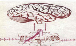
Receptors inform the brain of a patient about vertebrogene reflexive tonic and dystrophic changing of tissues.

Bending of the straight leg is stretching of ishiocrural and gluteale muscles.

If the reflexively contracted muscle resists this stretching it becomes a painful indicator of spreading of a painful zone.

Directing on a ishiocrural, popliteal or gluteale zone of pain it displays a zone, where palpation will discover tenderness.

If the patient informs about appearing that moment pain in the lumbar, it means that deformed part of the vertebral column contains a source of pain.

That means, that in the hypermobile segment of the column deformation of cinematic chain "leg - vertebral column" pain receptors are subjected by traumatism of hernia or other pathological structure.

If the defending muscle corset get a good nervous signaling, it protects the painful vertebral segment and at that moment the pain is absent.

Example 2.

The muscles of extremities are shortened while points of attaching are drawing together, whereas the back long muscles of the vertebral column are tensed, on the contrary, at the moment of their lengthening - when the body is bending forward for (not more that for 15-20 0 ).

When the patient is standing, in the state of rest these muscles are soft and the balance of the body is kept due to gravitation and muscles are not active.

If there is a painful vertebral segment pathological impulse causes sharp reflexive tension in the muscle.

That can be observed even in the state of quiet standing and doesn't disappear, when the body is bending more that for 20 0 , and in the state of standing only on one leg (the norm is that on the ipsilateral side paravertebral muscles are relaxing that moment).

Thus, in disposal of a vertebroneurologist there are methods of defining of quantitative and qualitative marks of reflexive reactions of the pathology of the vertebral column.

These marks allow to define not only the tonic and the character of the process but its dynamic development as well.

We hasn't touched here upon the problem of cure, which can be solved taking into consideration pathogenesis of mentioned here syndromology studied with the use of the developed scheme of investigation.

In conclusion there should be noted that owing to the well-known reasons the experience of Russian vertebroneurologists has not been yet claimed neither in Western Europe nor in the USA.

  Y. Y. Popelyanskiy  

01-04-2002


 Galina Popelianskaya
 
yakogalina@yahoo.com